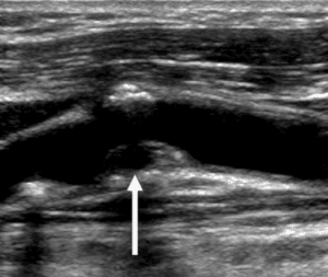
Ultrasound is a safe and painless tool that uses sound waves to produce images of our internal structures. Ultrasound is a wonderful imaging resource because it produces high quality diagnostic images without the use of radiation and it is noninvasive. Here at Windom Area Health, we perform many different kinds of ultrasounds, including vascular ultrasounds. Vascular ultrasound is when the technician is imaging any of the countless arteries or veins throughout the body. We are able to image the arteries and veins from your head to your toes, all in real-time.

Vascular disease can be a number of things, such as PAD (Peripheral Arterial Disease), DVT (Deep Venous Thrombosis) and CAD (Coronary Artery Disease), which involves the heart. These are all things that we are able to image on a daily basis here at Windom Area Health with the use of our in-house ultrasound, as well as mobile services. The risk factors for vascular disease include: smoking, diabetes, family history, heart disease, high cholesterol, and high blood pressure.
 We use ultrasound and specifically, Doppler, to assess the vasculature for disease in many different ways. There are many different imaging modes used via ultrasound. B-mode, or grayscale imaging, is used to visualize plaque buildup and clot formation in the arteries and veins. We are able to see the walls of these vessels to assess how severe the narrowing is. Think of your cardiovascular system like a hose. This is a very similar scenario to the plaque and clot formation process in your arteries and veins. If you were to press your hand down on the end of a hose, the water either moves very quickly, or completely halts. This is where Color Doppler and Pulsed Wave Doppler come into play.
With the use of Doppler, the blood flow is evaluated. Color Doppler shows how much blood is filling the artery or vein, and Pulsed Wave Doppler is evaluating the speed and characteristics of the blood flow. Pulsed Wave Doppler is similar to the waveform you would see on an EKG, when imaging an artery. We can see the systole and diastole of the cardiac cycle, although it does change in appearance depending on many factors. This is why Doppler is an extremely important resource for diagnosing disease. The waveform will look abnormal if there is a blockage somewhere in the artery or vein, and it aides the technician in finding that blockage. The technician then relays this vital information to the reading doctor, who writes the final report.

Ultrasound is vital in diagnosing and also after treatment of vascular disease. Here at Windom Area Health, we have a Registered Vascular Technologist that performs all types of vascular ultrasounds. We are also happy to work closely with Sanford Vascular Associates who provide services in our outreach center. Ask your doctor about vascular screenings if you have the risk factors for vascular disease!
We are currently hoping to replace our existing ultrasound machine with a newer model in the near future. This will allow us to better serve our patients with the latest technology.
Support your health and your local hospital by scheduling your ultrasounds at Windom Area Health today! Appointments can be scheduled Mondays-Friday 8:00-4:30 p.m. Call us today at 507-831-0670.
Blog Written By: Toriann Mortinsen, Ultrasound Tech, Registered Vascular Technologist

